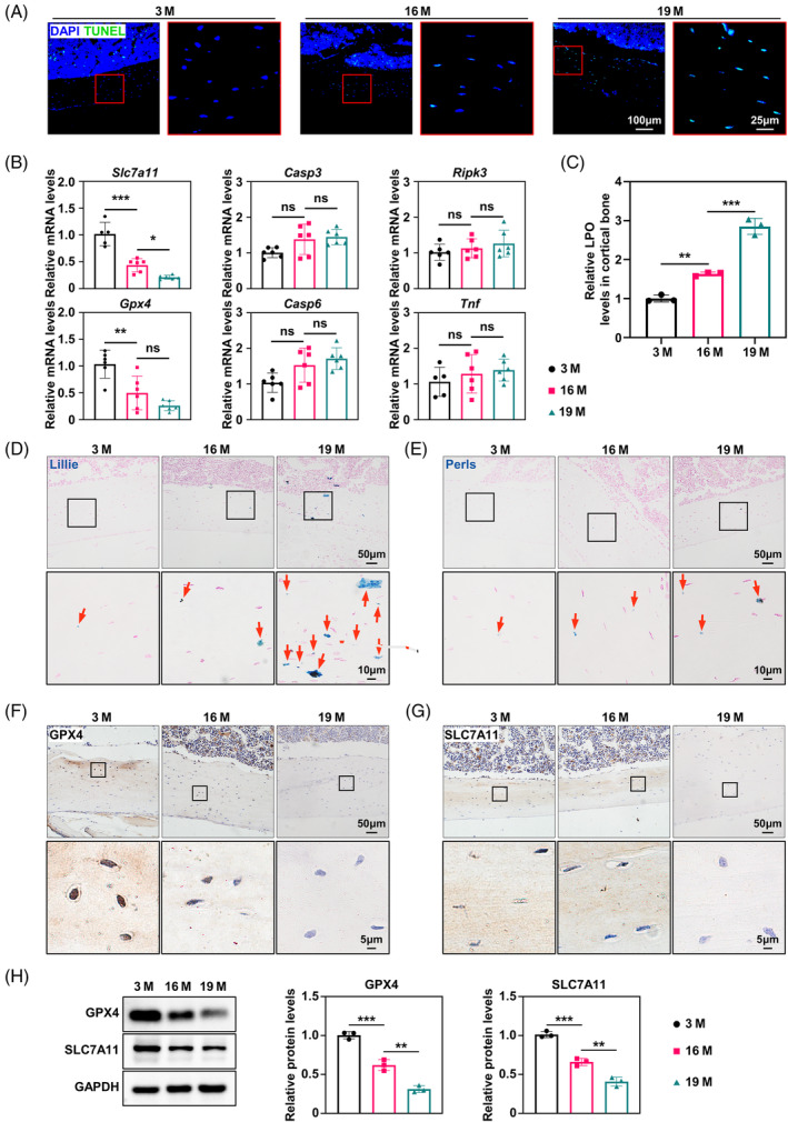
FIGURE 1.

Age‐related increases in cortical osteocyte death occur concurrently with a significant rise in ferroptosis. (A) Representative terminal deoxynucleotidyl transferase dUTP nick end labelling (TUNEL) staining of femur cortical bone from 3‐, 16‐ and 19‐month‐old mice (n = 3). (B) Quantitative reverse transcriptase PCR (qRT‐PCR) analysis of the levels of solute carrier family 7a member 11 (Slc7a11), glutathione peroxidase 4 (Gpx4), Casp3, Casp6, Ripk3 and Tnf in the femur cortical bone (n = 3). (C) Lipid peroxide colorimetric assay of femur cortical bone (n = 3). (D) Representative Lillie staining (blue) of the femur cortical bone, showing ferrous iron content in the cortical bone (n = 3). (E) Representative Perl's iron staining (blue) of the femur cortical bone, showing the ferric iron content in the cortical bone (n = 3). (F) Immunohistochemical staining for GPX4 in the femur cortical bone (n = 3). (G) Immunohistochemical staining for SLC7A11 in the femur cortical bone (n = 3). (H) Western blot analysis of the levels of GPX4 and SLC7A11 in the femur cortical bone, with quantitative data on the right (n = 3). M, months. *p < 0.05, **p < 0.01, ***p < 0.001.
