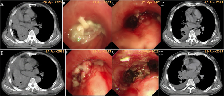
Figure 3.
Disease progression and treatment outcomes with multiple bronchoscopic interventions. (A) Chest CT showing right total atelectasis and mediastinal shift to the right; (B) Bronchoscopy showing complete obstruction of the right main bronchus lumen by a new growth; (C) Bronchoscopy after tumor resection showing tumor invasion of the right upper lobe; (D) Chest CT showing improvement in right atelectasis and mediastinal return to normal position after tumor resection; (E) Chest CT showing recurrence of right total atelectasis and right mediastinal displacement; (F) Bronchoscopy showing incomplete obstruction of the right main bronchus, right upper lobe bronchus by a new growth, and occlusion of the right middle bronchus; (G) Bronchoscopy after the third tumor resection; (H) Chest CT showing improvement in right atelectasis and mediastinal return to normal position after tumor resection.

