FIGURE 4.

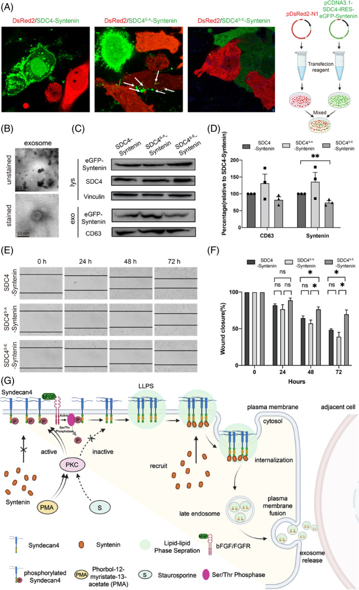
The dephosphorylation‐mimicking mutant SDC4S‐A‐syntenin increased the amount of syntenin in secreted exosomes and improved cell viability. (A) HeLa cells transfected with SDC4S‐A‐syntenin or SDC4S‐E‐syntenin and mixed with HeLa cells transduced with pDsRed2‐N1 for 72 h. Confocal microscopy showed that eGFP‐syntenin was detected in DsRed2‐labelled HeLa cells. (B) Transmission electron microscope (TEM) images showing the morphologies of SDC4‐syntenin exosomes. (C) Western blot showing that SDC4 phosphorylation decreased the amount of syntenin in secreted exosomes. (D) syntenin secretion from cells and in exosomes after SDC4 phosphorylation or dephosphorylation; n = 3 biologically independent samples, and the data are presented as the mean values ± SEMs. Comparisons between groups were performed via two‐tailed unpaired t test (**p < 0.01). (E) Percentage of the wound closed after SDC4‐syntenin, SDC4S‐A‐syntenin or SDC4S‐E‐Sy overexpression. Micrographs were taken immediately after wounding and 24, 48, and 72 h after the introduction of a wound. Black dashed lines denote wound edges. (F) Values significantly different from controls are indicated with an asterisk (*p < 0.05). The graphs show the means (with SEMs). (G) Schematic illustration showing the proposed model in which SDC4 phase separation and syntenin on the plasma membrane regulate the exosomes biogenesis and intercellular signalling activity.
