Figure 4.

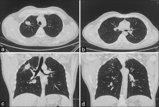
Follow-up CT of the chest taken after 3 months (a and b) Axial section (c and d) Coronal section showing showing near complete resolution with fibro nodular opacity in right upper lobe

Follow-up CT of the chest taken after 3 months (a and b) Axial section (c and d) Coronal section showing showing near complete resolution with fibro nodular opacity in right upper lobe