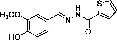
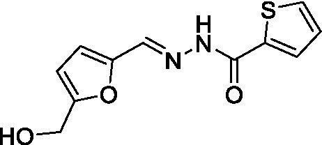
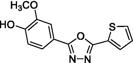
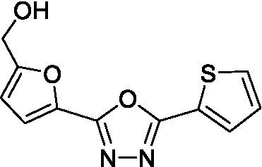
Table 1.
Binding parameters for TcBDF3 by thermal shift and fluorescence polarization*.
| Compound | Structure | Kd ± SD DSF (μM) | IC50 ± SD FP |
|---|---|---|---|
| A1B4 |

|
1.7 ± 0.2 (García et al., 2018) | 2.4 ± 0.3 |
| A5B4 |

|
136 ± 2.4 | n.d. |
| A1B4c |

|
4 ± 0.3 | 8.4 ± 0.5 |
| A5B4c |

|
4.8 ± 0.2 | 10.5 ± 0.9 |
*Kd for A1B4 by DSF corresponds to that already published (García et al., 2018). The results are expressed with the SD obtained from three independent experiments. Each experiment was performed in triplicates. CC50, cytotoxic concentration; DSF, differential fluorescence scanning; FP, fluorescence polarization; n.d., not determined; SD, standard deviation.
