Fig. 1.
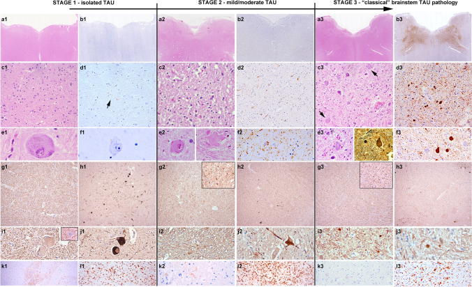
Histological findings at the different stages of pathology. Representative histological images of the neuropathological features of anti-IgLON5 disease at different grades of severity. a1–f1, a2–f2 and a3–f3: First row: HE and Tau immunohistochemistry at the level of the medulla oblongata. At low magnification (a1–b3), extensive tau pathology can be clearly observed in the tegmentum at stage 3 (b3), but not at stage 1 (b1) and barely at stage 2 (b2). Second row: at higher magnification (c1–d3), Tau pathology is already visible at stage 2 (d2) in form of neuropil threads and pretangles, while at stage 1 isolated delicate threads are visible (d1, arrow). In contrast, HE-stained sections in stage 1 (c1) and stage 2 (c2) already show reactive changes in the reticular formation. In addition, some enlarged neurons are observed at stage 1 and stage 2 (e1, e2), that are tau negative, while at stage 3, they are already containing tau-positive neurofibrillary tangles, but are overall reduced in numbers. These enlarged neurons show a slightly increased immunoreactivity for phosphorylated (SMI31; g1, i1) and particularly of non-phosphorylated (SMI32; h1, j1) neurofilaments, and have a regular axonal density and morphology. In stage 2 and stage 3, the number of enlarged neurons decreases (h2, j2 and h3, j3) but an increase in axonal spheroids is detected (i2, i3). k1, k2, k3: Immmunohistochemistry for IgG4 in the brainstem shows focally marked deposits at stage 1 (k1), mild deposits in single cases at stage 2 (k2) and no deposits at stage 3 (k3). i1, i2, i3: different grades and states of microglial activation at different disease stages (HLA-DR immunohistochemistry)

