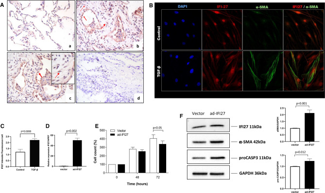
Fig. 6.
IFI27 overexpression reduces the cell count over time and induces activation of lung fibroblasts. (A) IFI27 immunostaining in lung tissues. (a) Lung tissue from control donor. (b-c) The positive signal for the IFI27 protein was in cells with epithelial (solid arrow) and mesenchymal morphology (dotted arrow) in HP. (d) Immunostaining of HP lung tissue where the primary antibody was omitted. (B) Immunofluorescent localization of IFI27 and αSMA. (C) Quantification of IFI27 means fluorescence intensity in (B). (D) qPCR analysis of IFI27 expression in transduced fibroblasts (E) CyQuant assay in normal lung fibroblasts (NHLF and HPF) transduced with the empty adenoX-DS-Red-Express (vector) and vector coding the full-length IFI27 (ad-IFI27). Two independent experiments in quadruplicate were done in two control cell lines. (F) Representative immunoblots of IFI27, pro-caspase 3, and αSMA. The side graph represents the densitometric analysis of three independent experiments of relative values of target proteins compared to GAPDH as a loading control. The images were extracted from the original blots to show the bands of interest. RNA and protein samples were collected 48 h after transduction.

