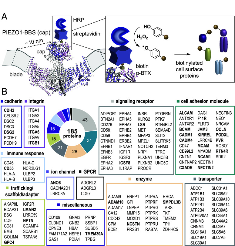
Fig. 1.
Proteomic mapping of the PIEZO1 surface interactome. (A) Side view of the PIEZO1 trimeric structure (PDB: 6BPZ) in the plasma membrane, illustrating the PIEZO1-BBS (cap) construct bound to a cartoon of α-BTX-biotin and a streptavidin-HRP conjugate for biotinylation of extracellularly exposed proteins within ~20 nm of the cap. The distance between the top of the cap domain and end of the extracellular distal blade is ~10 nm, based on other resolved structures (black dotted line). For clarity, the distal blade repeats are not well resolved in the 6BPZ structure and are represented by gray dotted lines within the plasma membrane. (B) Set of proteins found by proximity labeling and MS-based proteomics to reside in the vicinity of PIEZO1-BBS (cap), annotated by functional classifications. Bolded proteins were selected for follow-up functional screening (Fig. 2A).

