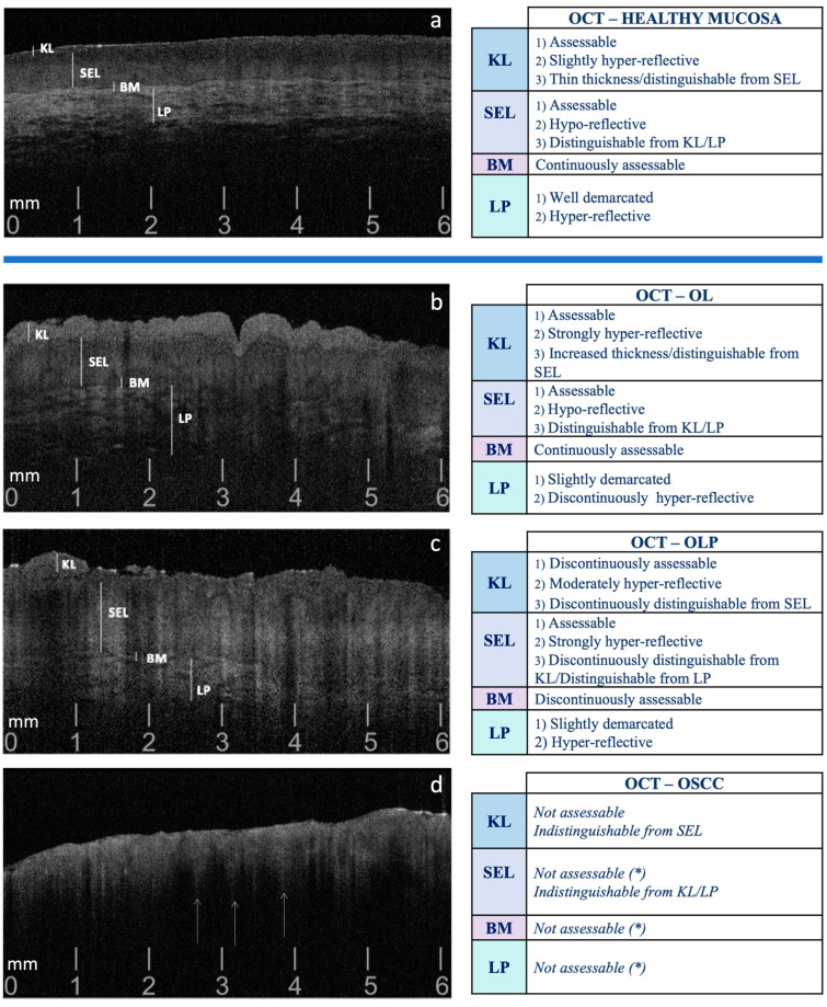
Figure 1.
Guiding pattern criteria for the selection of OCT scans, comparing the patterns observed in healthy oral mucosa (a) with those in OL (b), OLP (c), and OSCC (d). OL: Oral Leukoplakia; OLP: Oral Lichen Planus; OSCC: Oral Squamous Cell Carcinoma; KL: Keratinized Layer; SEL: Stratified Epithelial Layer; BM: Basement Membrane; LP: Lamina Propria. (*) Presence of ‘icicle-like’ structures: hyper-reflective conical configurations that extend from the superficial cellular layers (SEL) to the deeper ones (BM and LP), commonly reported in OSCC, suggestive of neoplastic intra/sub-epithelial infiltration (indicated by white arrows ↑) [19].

