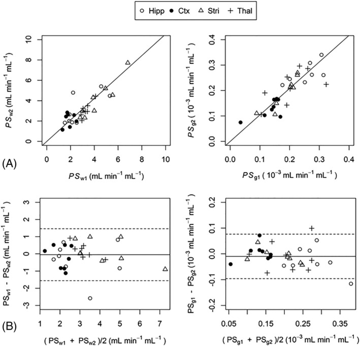
FIGURE 2.

Correlation (A) and Bland–Altman (B) plots showing the agreement between regional scan‐rescan measurements of PS w and PS g . PS w and PS g had coefficient of determination values (R 2) of 0.82 (p < 10−12) and 0.96 (p < 10−16), respectively. Solid lines in the Bland‐Altman plots show the mean difference between scan 1 and scan 2. Dashed lines show the limits of agreement within which 95% of scan‐rescan differences lie. Hipp, hippocampus; Ctx, cortex; Stri, striatum; Thal, thalamus
