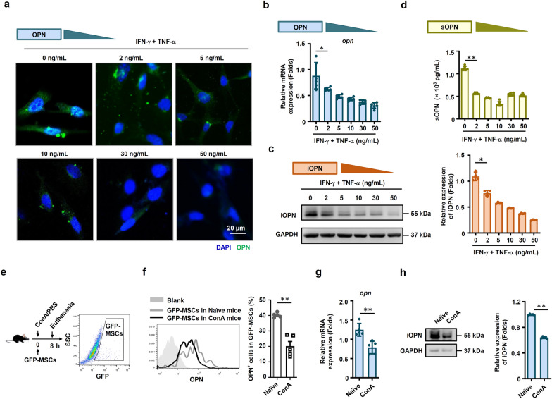
Fig. 1.
OPN expression was downregulated in MSCs under inflammatory conditions. a MSCs were treated with TNF-α plus IFN-γ at the indicated concentration for 24 h. Images were collected by a Zeiss fluorescence microscope. Scale bar: 20 μm. b and c Murine MSCs were treated with IFN-γ plus TNF-α at the indicated concentration for 24 h, and mRNA and protein of MSCs were collected. OPN expression was determined at the mRNA and protein levels by quantitative real-time PCR and immunoblotting analysis. Full-length blots are presented in Additional file 1: Fig. 1c. d sOPN in the culture medium of MSCs treated with IFN-γ plus TNF-α at the indicated concentration for 24 h was tested by enzyme-linked immunosorbent assay. e–h MSCs with CFSE staining were administered intravenously to mice with ConA-induced liver damage. The mice were euthanized after 8 h, and the mononuclear cells in mouse lungs were collected. OPN expression in GFP+ mononuclear cells was evaluated by flow cytometry (f), quantitative real-time PCR (g) and immunoblotting analysis (h) (naïve: n = 5, ConA: n = 5). Full-length blots are presented in Additional file 1: Fig. 1h. The results are representative of three to six independent experiments. Values are shown as the mean ± SEM and statistical significance is indicated as *P < 0.05 and **P < 0.01

