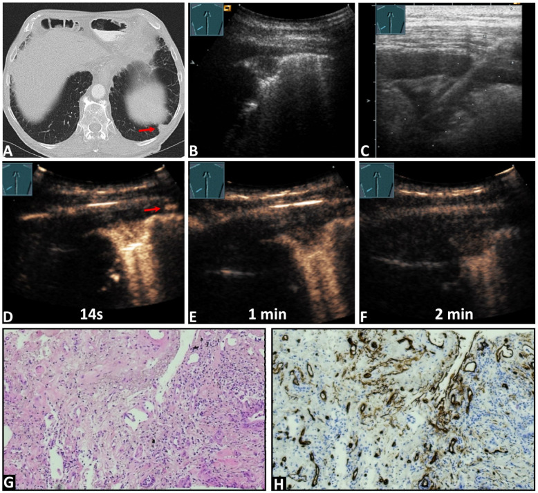
Figure 3.
An 84-year-old male patient with known pancreatic carcinoma and a pulmonary nodule on the (A) computed tomography (arrow) (courtesy of Prof. Dr. Andreas H. Mahnken, Department of Radiology, University Hospital Marburg) and (B,C) B-mode ultrasound. (C) An ultrasound-guided 18G core needle biopsy of the lung lesion was performed, and histopathological examination confirmed the diagnosis of metastatic pancreatic carcinoma. On contrast-enhanced ultrasound, the lesion demonstrated (D) an enhancement simultaneous with the arrival of the contrast agent in the intercostal artery (arrows), and a marked and homogeneous pattern of enhancement with (E,F) an early decrease of enhancement. (G) The tissue sample (HE staining) showed the infiltrates of a ductal adenocarcinoma consistent with a metastasis of the previously known pancreatic carcinoma. (H) immunohistochemical staining with CD34 was performed, revealing a chaotic pattern consistent with bronchial-arterial neoangiogenesis (×100).

