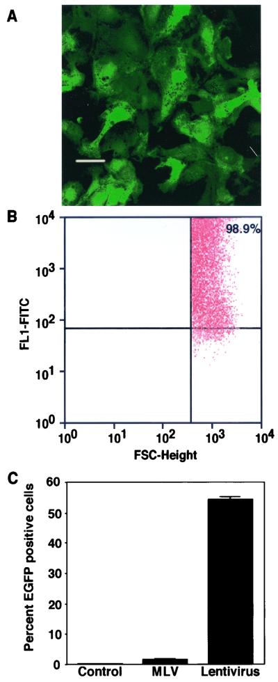
FIG. 2.
Transduction of AEC with VSV-G-pseudotyped lentivirus vectors. (A) AEC in primary culture on chamber slides were transduced with VSV-G-pseudotyped lentivirus vectors at an MOI of 10. Confocal microscopy from a representative experiment at 3 days posttransduction demonstrates strong expression of EGFP in the majority of cells. Bar, 20 μm. (B) AEC grown on plastic tissue culture dishes were transduced with VSV-G-pseudotyped lentivirus vectors. Representative FACS analysis at 72 h after infection demonstrates 99% EGFP-positive cells at an MOI of 50. (C) Transduction of AEC grown on plastic is ∼30-fold greater for VSV-G-pseudotyped lentivirus vectors than VSV-G-pseudotyped MLV-based vectors at a comparable titer.

