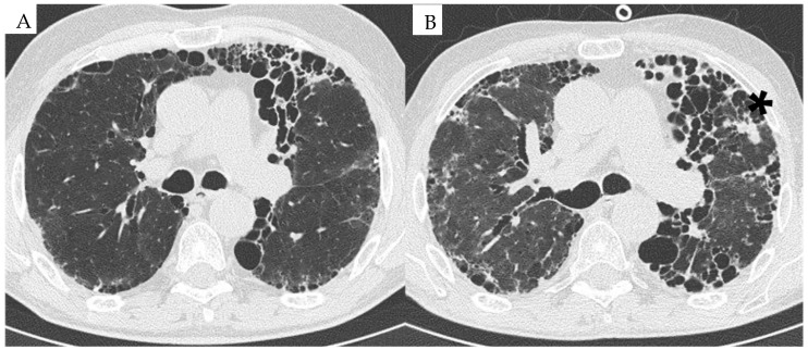
Figure 1.
HRCT image of a patient with RA-ILD at baseline in 2015 (A) and follow-up in 2023 (B). (A) UIP pattern associated with RA associated with reticulations and mild ground-glass opacities. (B) Progression of the UIP pattern with the occurrence of a neoplastic lung nodule in the left upper lobe (*).

