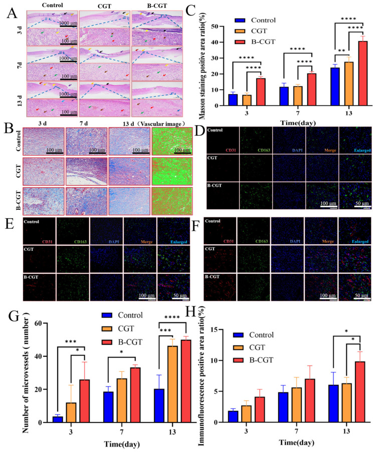
Figure 8.
The effect of B-CGT on wound healing. (A) The morphological changes of the skin layer at different time points. (B) Masson staining analysis. (C) Masson positive area ratio. (D) Fluorescence double-labeled detection of wound tissue on the third day. (E) Fluorescent double-labeled detection of wound tissue on the seventh day. (F) Fluorescence double-labeled detection of wound tissue on the 13th day. (G) The number of microvessels in the wound tissue at different time points. (H) Immunofluorescence-positive area ratio. * p < 0.05, ** p < 0.01, *** p < 0.001, **** p < 0.0001.

