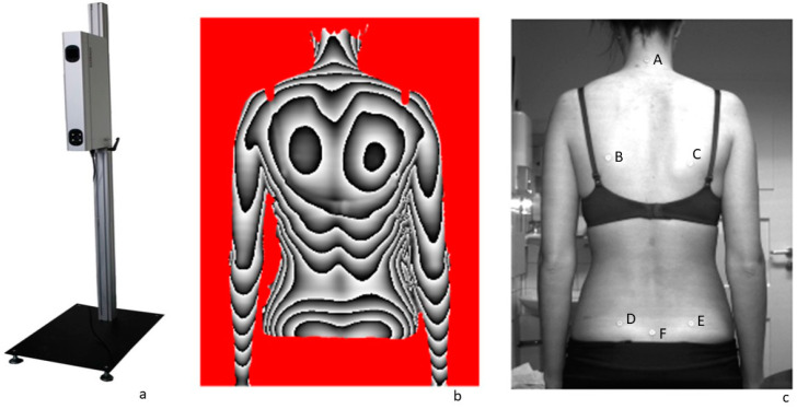
Figure 1.
(a) MiniRot Combi back scanner (ABW GmbH, Frickenhausen/Germany), (b) three-dimensional phase picture of the back, (c) illustration of the exact marker position on the back: A: vertebra prominens (7th cervical vertebra), B: left lower scapular angle, C: right lower scapular angle, D: left spina iliaca posterior superior (SIPS), E: right spina iliaca posterior superior (SIPS), F: sacrum point (cranial beginning of the gluteal cleft).

