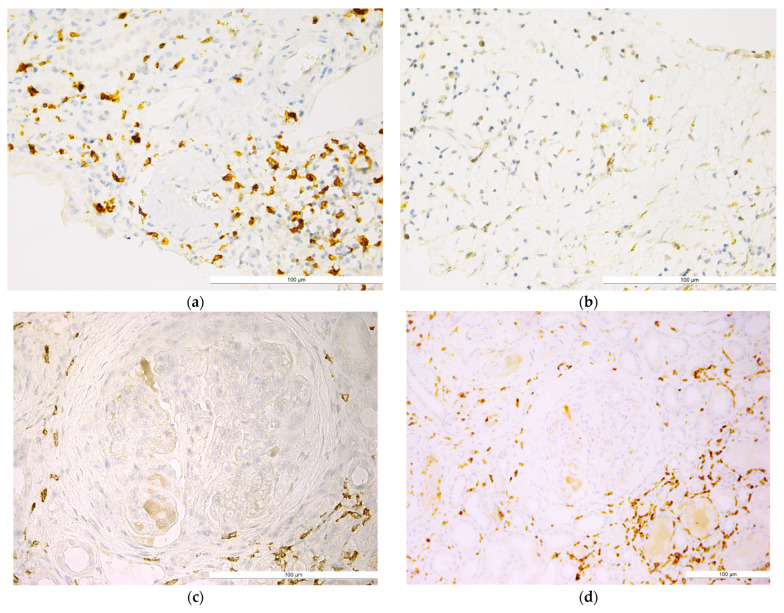
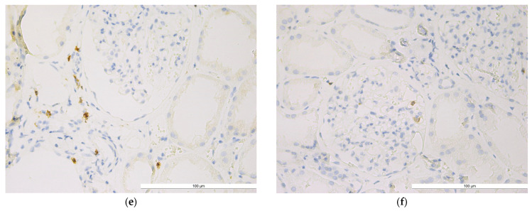
Figure 1.
Lupus nephritis with (a) well-represented periglomerular and interstitial CD4+ T lymphocytes (IHC, anti-CD4, ×40); (b) CD4+ T lymphocytes in the renal interstitial space in an area of tubular atrophy (IHC, anti-CD4, ×40); (c) CD4+ T lymphocytes with concentric periglomerular localization (IHC, anti-CD4, ×40); (d) CD4+ T lymphocyte population in the periglomerular and interstitial areas (IHC, anti-CD4, ×20); (e) poorly represented periglomerular and interstitial CD4+ T lymphocyte population (IHC, anti-CD4, ×40); (f) isolated CD4+ T lymphocytes present in the intraglomerular area (IHC, anti-CD4, ×40). The bars indicate a size of 100 µm.


