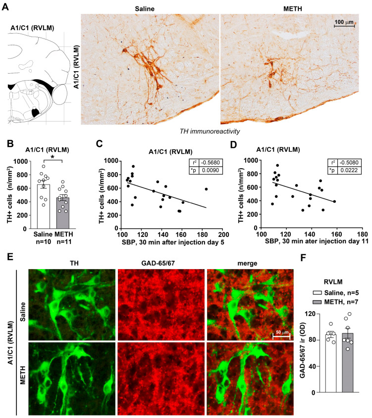
Figure 3.
Within the RVLM, repeated METH administration reduces TH immunopositive cells without affecting GAD immunopositive cells. Immunohistochemical analysis for the catecholamine marker TH was carried out from dissected brains of mice subjected to repeated injections of saline or METH (5 mg/kg, i.p., for 5 days) and then sacrificed 60 min after the METH/saline challenge, which was carried out on day 11. Representative images of TH-positive neurons within A1/C1 in the RVLM of mice treated with saline or METH are shown in (A). The graph in (B) shows TH-positive cell density (n/mm2) counted in saline- and METH-treated mice. Values are the means ± S.E.M. * p < 0.05 (unpaired two-tailed Student’s t-test). The correlation analyses between TH-positive cell density within A1/C1 in the RVLM and SBP are shown in (C,D), respectively. These correlation analyses were carried out considering SBP values that were measured at 30 min following the fifth saline/METH injection or following the METH/saline challenge. * p < 0.05, Pearson correlation test. (E) Representative images of double immunostaining for TH and GAD in the RVLM of mice following repeated injections of saline or METH (5 mg/kg, i.p., for 5 days). These mice were sacrificed 60 min following the METH-/saline challenge, carried out on day 11. The densitometry (OD: Optical Density) of GAD65/67 immunoreactivity (ir) is shown in (F). Values are the means ± S.E.M. * p < 0.05 (two-tailed Mann-Whitney test).

