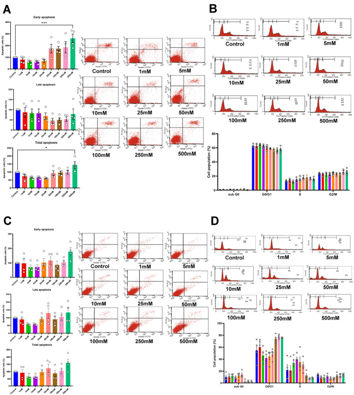
Figure 3.
Apoptotic cell analysis using flow cytometry of HSC-4 (A) and HSC-7 (C) after 72 h of betaine treatments. Cell cycle analysis of HSC-4 (B) and HSC-7 (D) after betaine treatment for 72 h. A normal culture medium was used as a control. Bar and asterisks indicate a significant difference. Each small circle represents an individual sample. The data are shown in mean ± SE, n = 4.

