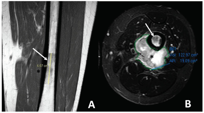
Figure 6.
Soft tissue spreading in conventional high-grade osteosarcoma affecting the left femoral bone diaphysis in a 20-year-old male. (A) Coronal T1-weighted imaging (WI) showing the irregular intra-medullary involvement with a low signal intensity (white arrow) and a soft-tissue extension (black asterisk). (B) Axial T2-WI with fat suppression showing ill-defined soft-tissue spreading with high, heterogeneous signal intensities on T2-WI. Of note, the extra-osseous was automatically segmented (green line) for a further volumetric follow-up during neoadjuvant treatments.

