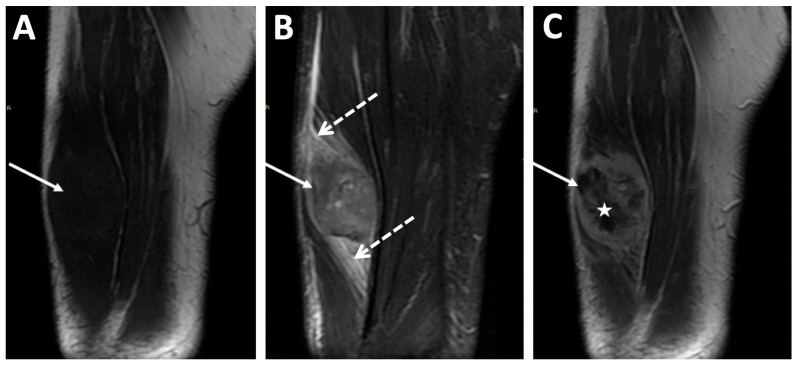
Figure 8.
MRI of a high-grade extraskeletal osteosarcoma of the right thigh diagnosed in an 83-year-old male. (A) Coronal T1-weighted imaging (WI), (B) fat-suppressed coronal T2-WI, and (C) coronal T1-WI with gadolinium chelate injection. The tumor was seated deep in the anterior compartment of the thigh (arrows) and demonstrated a peritumoral edema (dashed arrows above and below the tumor) and inhomogeneous enhancement with internal necrosis (star).

