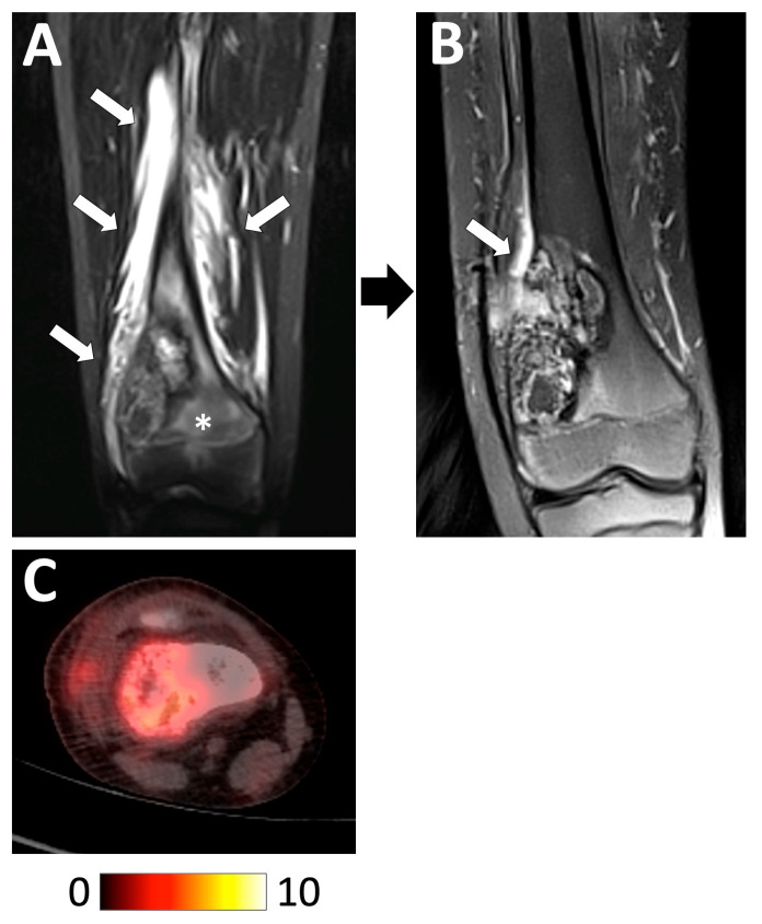
Figure 11.
Examples of radiological features associated with a good histologic response. A 13 year-old boy diagnosed with a high-grade osteoblastic osteosarcoma of the distal right femoral bone was treated with perioperative chemotherapy. (A) Initial coronal STIR T2-weighted imaging showed the tumor with intramedullary edema (white asterisk) and extensive peritumoral edema in the surrounding tissues (white arrows). (B) At the end of chemotherapy, the tumor contours were better defined with a marked decrease in the intramedullary and soft-tissue edema. (C) Initial 18F-FDG PET/CT showing a moderately hypermetabolic tumor (baseline SUVmax = 5.65). The pathological analysis of the curative surgical resection revealed a good response (necrosis rate = 94.5%) and R0 margins. The patient is still alive without relapse 8 years later.

