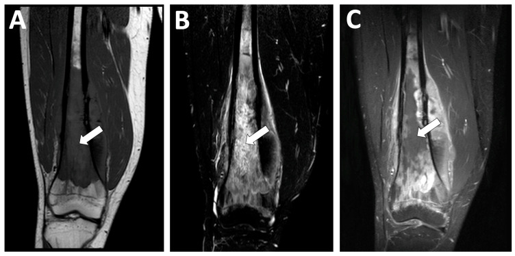
Figure 12.
Examples of radiological features associated with a poor histologic response. A 15 year-old male presented with a high-grade osteoblastic osteosarcoma of the distal femoral bone. This tumor was characterized by a longest diameter of 16 cm, a volume > 150 mL, as well as central necrosis > 50% of the tumor volume (white arrows) with an intermediate signal intensity on (A) coronal T1-weighted imaging (WI), a high signal intensity in STIR (B), and no contrast enhancement on (C) fat-suppressed contrast-enhanced T1-WI. After completing neoadjuvant chemotherapy, a pathological analysis of the surgical specimen demonstrated a poor response (necrosis rate = 80%) and R0 margins. However, the patient is still alive without relapse 10 years later.

