Figure 1.
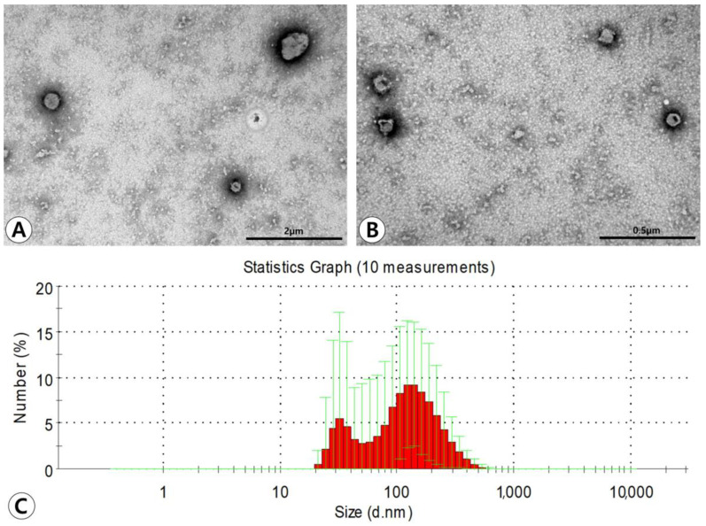
Characterization of adipose stem cell (ASC)-derived extracellular vesicles (EVs). (A,B) Transmission electron microscopy demonstrated that ASC-derived EVs have lipid bilayers and spherical shape (original magnification ×50,000 and ×80,000, respectively). (C) The average size of the EVs measured by dynamic light scattering was 130.3 ± 65.7 nm in diameter. The majority of particles isolated from the ultracentrifugation-concentrated EV pellets were in a size range of approximately 100–200 nm.

