FIG. 2.
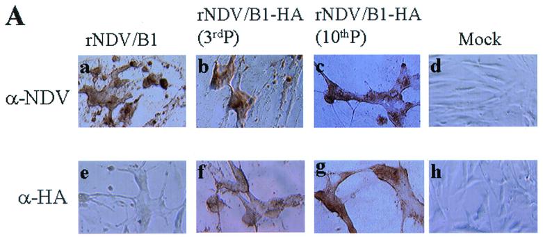
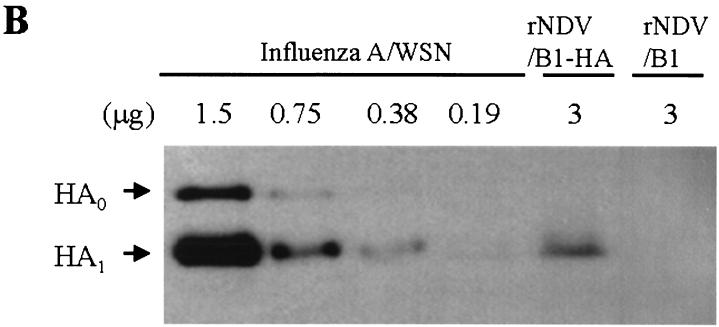
Detection of the HA protein on infected cells and in purified virions. (A) rNDV/B1- or rNDV/B1-HA-infected cells were fixed with 1% paraformaldehyde at day 2 postinfection, and cells were used for immunostaining analysis. NDV protein expression on the surfaces of cells infected with rNDV/B1 (a) or rNDV/B1-HA passaged in eggs at a low multiplicity of infection three times (3rdP) (b) or 10 times (10thP) (c) was analyzed by using mouse anti-NDV serum. HA expression on the cell surfaces of rNDV/B1 (e)- or rNDV/B1-HA (f and g)-infected cells was analyzed by using anti-HA monoclonal antibody (2G9). Mock-infected cells were also analyzed as a control (d and h). (B) rNDV/B1 and rNDV/B1-HA were purified from allantoic fluids of infected embryonated chicken eggs. Influenza A/WSN/33 virus was purified from MDBK cell culture supernatants as a control. Serial twofold dilutions of A/WSN/33 viral proteins (1.5 to 0.19 μg) and 3 μg of rNDV/B1-HA or rNDV/B1 viral proteins were separated on a sodium dodecyl sulfate–10% polyacrylamide gel. The gel was transferred to a nitrocellulose membrane, and the HA protein was detected by chemiluminescence using a mixture of anti-HA monoclonal antibodies and an anti-mouse IgG peroxidase-labeled antibody (DAKO). HA0 and HA1 indicate uncleaved and cleaved HA protein, respectively.


