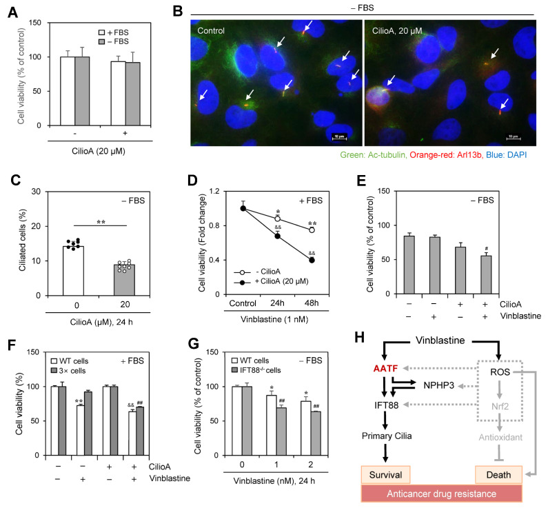
Figure 8.
Inhibition of primary cilium formation by ciliobrevin A (CilioA) treatment enhanced vinblastine (VBL)-mediated cancer cell death. (A–C) HeLa cells were incubated in the absence or presence of CilioA. Cell viability was measured by CellTiter Glo luminescent assay (A). Cells incubated under serum-starved (SD) conditions were fixed and stained with antibodies against Ac-tubulin (green) and Arl13b (orange-red). The nucleus was stained with DAPI (blue). The primary cilium was observed and photographed at 400× or 1000× magnification with 40× or 100× objectives, respectively under a fluorescence microscope. The image with primary cilia at 1000× magnification is representative of ≈30 pictures. Processing (such as changing brightness and contrast) is applied equally to control across the entire image. White arrows indicated primary cilia (B). The ciliated HeLa cells (n > 500 cells) in the absence (white) or presence (grey) of CilioA were counted (C). (D–G) Wildtype (WT), 3× HeLa cells treated with VBL repeatedly three times or PC-deficient IFT88−/− A375 cells were incubated with 1 nM VBL in the presence (D,F) or absence (E,G) of FBS for 24 h (D–G) or 48 h (D). Cell viability was measured by the trypan blue exclusion assay (D–F) or CellTiter Glo luminescent assay (G). Data were representative of four experiments. Data in the bar (A,C,E–G) or line (D) graphs represent the means ± SEM. * p < 0.05, ** p < 0.01; significantly different from the CilioA-untreated (C) or VBL-untreated (D,F,G) control group. && p < 0.01; significantly different from CilioA-untreated and VBL-treated group in the presence of FBS (D,F). # p < 0.05, ## p < 0.01; significantly different from CilioA-untreated and VBL-treated WT cell group in the absence of FBS (E) or 3× cell group in the presence of FBS (F) or VBL-treated WT cell group at each concentration (G). (H) Schematic mechanism for primary cilia formation by VBL-induced IFT88 through AATF and NPHP3 expression to regulate cancer cell survival and death. AATF regulated NPHP3 and IFT88 expression and primary cilium formation in HeLa human cervical cancer cells (solid line). ROS also upregulates NPHP3 expression through Nrf2 and unknown factors (grey dotted lines). Our findings were indicated by black solid lines. The pathway already known was indicated by grey solid lines.

