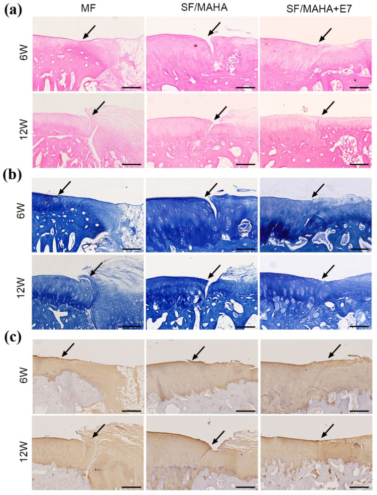
Figure 6.
Histological assessments of neocartilage formation in the injured area. (a) Hematoxylin–eosin staining. (b) Toluidine blue staining. (c) Immunohistochemical staining for Col II. (the arrows indicate the margins of the normal cartilage and repaired cartilage) (Scale bar = 200 μm) MAHA, methacrylated hyaluronic acid. SF, silk fibroin.

