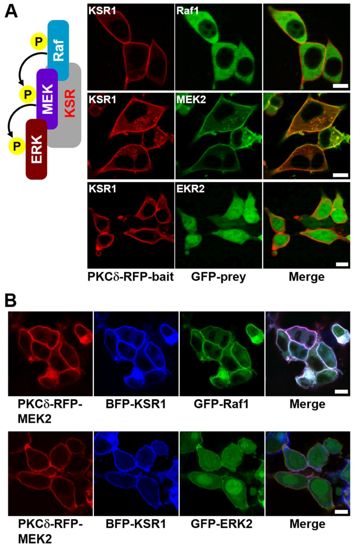
Figure 4.
Confocal images of the Raf–MEK–ERK signaling cascade in living cells with KSR1 scaffold protein. (A) HEK-293T cells were transiently co-transfected with PKCδ–mRFP–KSR1 and each of eGFP–Raf1, eGFP–MEK2, and eGFP–ERK2. The cells were then serum-starved, stimulated with EGF, and treated with PMA. KSR1 translocation to the plasma membrane was observed, with or without interaction with Raf1, MEK2, or ERK2, as indicated (top, middle, and bottom rows, respectively). (B) HEK-293T cells were transiently co-transfected with either PKCδ–mRFP–MEK2/TagBFP–KSR1/eGFP–Raf1 or PKCδ–mRFP–MEK2/TagBFP–KSR1/eGFP–ERK2. The cells were then serum-starved, stimulated with EGF, and treated with PMA. The ternary protein complexes, both Raf1/KSR1/MEK2 and MEK2/KSR1/ERK2, were co-translocated to the plasma membrane. The images were acquired 3–5 min after PMA treatment. The scale bar is 10 μm.

