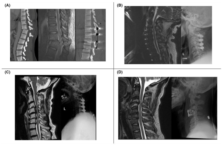
Figure 1.
(A) Patient with lower extremity motor and sensory loss after MVC (ASIA A); CT (left) and sagittal T2 MRI (middle) show T11–12 fracture dislocation; postoperative CT shows improved alignment (right). (B) Patient with intact sensation but no motor function after fall (ASIA B); preoperative MRI (left) shows C3–4 stenoses and cord contusion; postoperative decompression and fusion from C2–5 X-ray (right). (C) Patient with 2/5 hand weakness and intact sensation (ASIA C) after fall; MRI with spinal cord contusion at C4–6 (left); postoperative X-ray with instrumented decompression and fusion (right). (D) Patient with hand paresthesia and intact motor function after fall (ASIA D); MRI with traumatic disc herniation at C3–4 (left); postoperative C3–4 anterior cervical discectomy and fusion X-ray (right).

