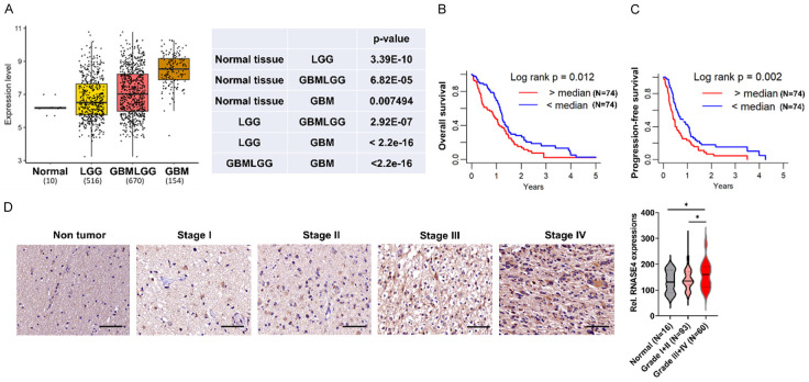
Figure 1.
RNASE4 expression correlates positively with glioblastoma malignancy grade. A. TCGA analysis illustrating a progressive increase in RNASE4 mRNA levels from normal brain tissue (n=10) to lower-grade glioma (LGG, n=516), a combination of lower-grade glioma and glioblastoma (GBMLGG, n=670) and glioblastoma (GBM, n=154). B, C. Kaplan-Meier plots depicting significantly reduced overall and progression-free survival in TCGA GBM patients with high versus low RNASE4 expression. D. Immunohistochemistry images and quantification revealing elevated RNASE4 protein levels in high-grade (III-IV) compared to low-grade (I-II) glioma tissues and normal brain. Scale bar, 50 μm. The right panel shows RNASE4 immunohistochemistry scores, indicating a progressive upregulation with advancing tumor grade. Data are presented as mean ± SEM. *P<0.05.

