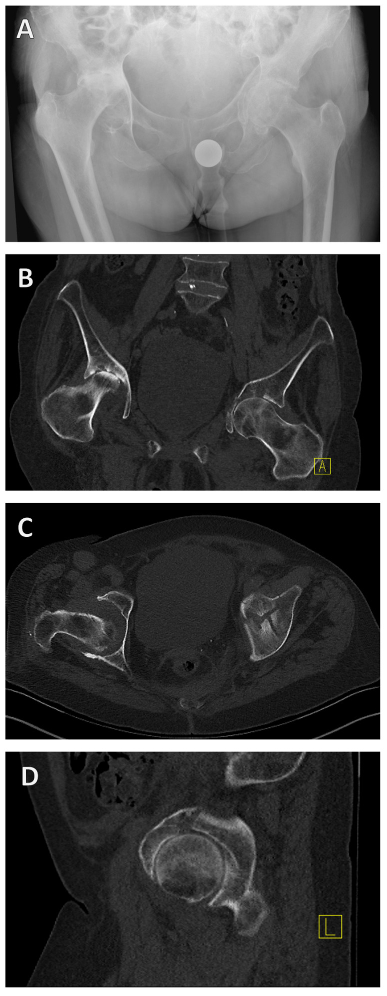
Figure 3.
X-ray and CT diagnosis of a geriatric acetabular fracture. Initially, the left-sided acetabular fracture can be recognized in the anterior–posterior radiograph of the pelvis (A). A CT scan was added for fracture classification and preoperative planning (B–D). It revealed a T-shape fracture (B–D).

