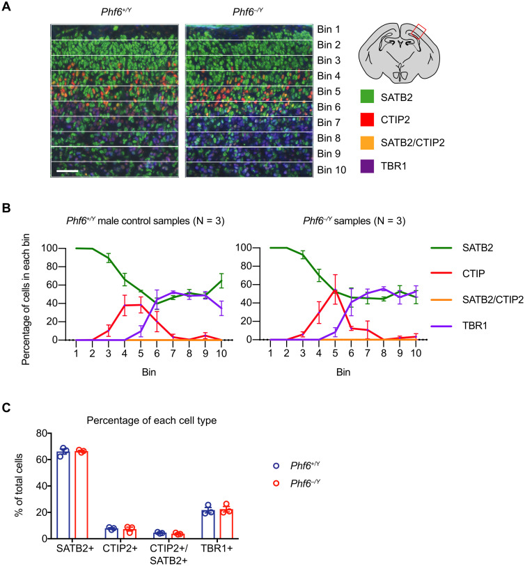
Fig 4. Germline Phf6 deletion does not cause a cortical lamination defect.
(A) Representative images of cortical layer staining using SATB2, CTIP2, and TBR1 showing the developing Phf6+/Y and Phf6–/Y E18.5 parietal cortex divided into 10 pial to ventricular bins. Scale bar = 50 μm. (B) Percentage of cells expressing the marker proteins in each bin for three mice per genotype. No significant differences between genotypes were observed. (C) Percentage of cells of each type across the entire thickness of the cortex. No significant differences between genotypes were observed. N = 3 Phf6+/Y and 3 Phf6–/Y foetuses. Data are presented as mean ± sem and were analysed by two-way ANOVA with Šídák’s multiple comparisons test. Circles (C) represent individual mouse foetuses. Related data are shown in S8 and S9 Figs; assessment of neurite outgrowths in cultured cortical neurons is displayed in S10 Fig.

