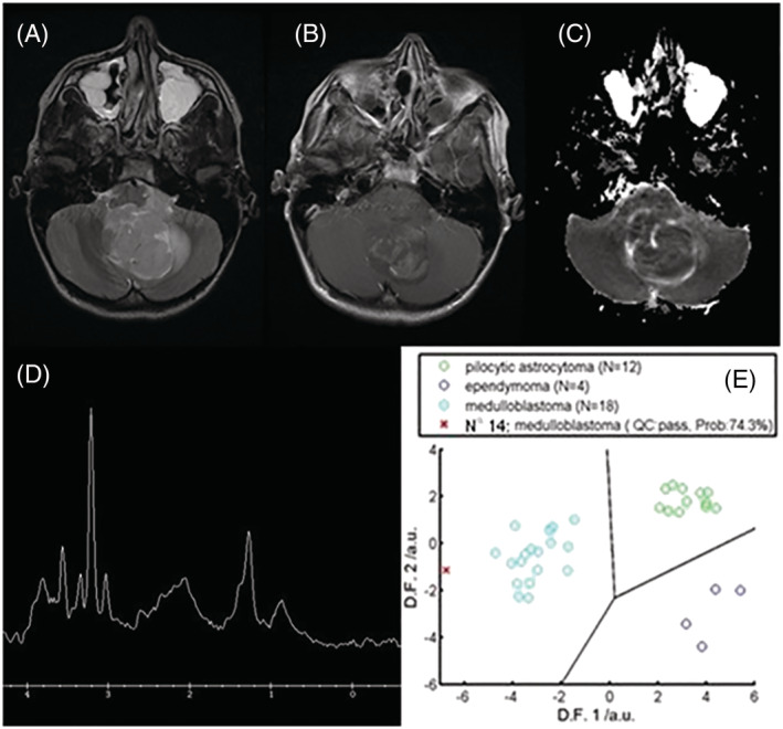
FIGURE 6.

A 13‐year‐old boy presented with headaches and vomiting and was found to have a 4.2 x 3.6 x 2.4 cm heterogeneous mass lesion in the right cerebellar hemisphere on MRI. The lesion contained multiple cystic areas and prominent vessels and demonstrated restricted diffusion. MRS demonstrated a high Cho peak, low Ins and the presence of taurine, a pattern observed in medulloblastoma. Histology confirmed a diagnosis of medulloblastoma 8 days after surgical resection. The figure shows (A) T2‐weighted MRI, (B) postcontrast T1‐weighted MRI, (C) an apparent diffusion coefficient (ADC) map, (D) MRS processed by scanner and (E) DSS output. The radiologists' assigned certainty for the correct diagnosis increased from 50% to 80%, from 45% to 75%, and from 40% to 60% for radiologist 1, 2 and 3, respectively, with the inclusion of MRS and DSS
