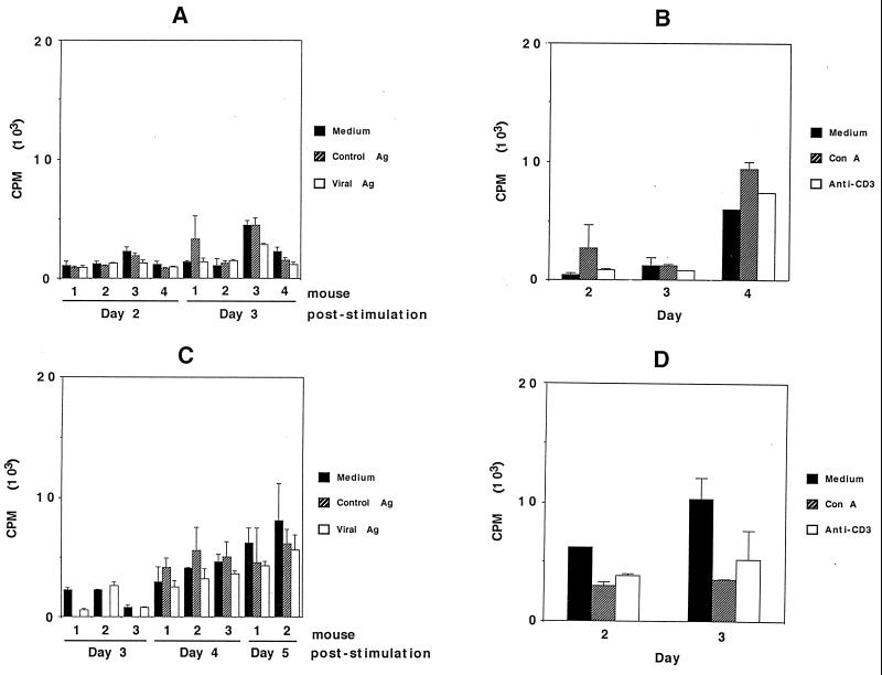
FIG. 5.
In vitro proliferation assay of brain-associated lymphocytes from parental (A and B) and GKO (C and D) mice which had been immunized and virus challenged. (A) Lymphocytes were isolated on day 5 postchallenge. The cells were stimulated with viral antigen (Viral Ag), SW-13 cell antigen (Control Ag), or medium alone for 2 and 3 days. (B) Cells harvested from three mice on day 5 postchallenge were pooled and tested for stimulation by ConA or anti-CD3 antibody after 2 to 4 days. (C) Cells were harvested on day 5 postchallenge and stimulated with viral antigen, control antigen, or medium. The results after 3 to 5 days of stimulation are shown. (D) Cells from three mice on day 5 postchallenge were pooled and stimulated with ConA or anti-CD3 antibody, and proliferation was measured on day 2 or 3 following stimulation. The error bars indicate standard deviations.

