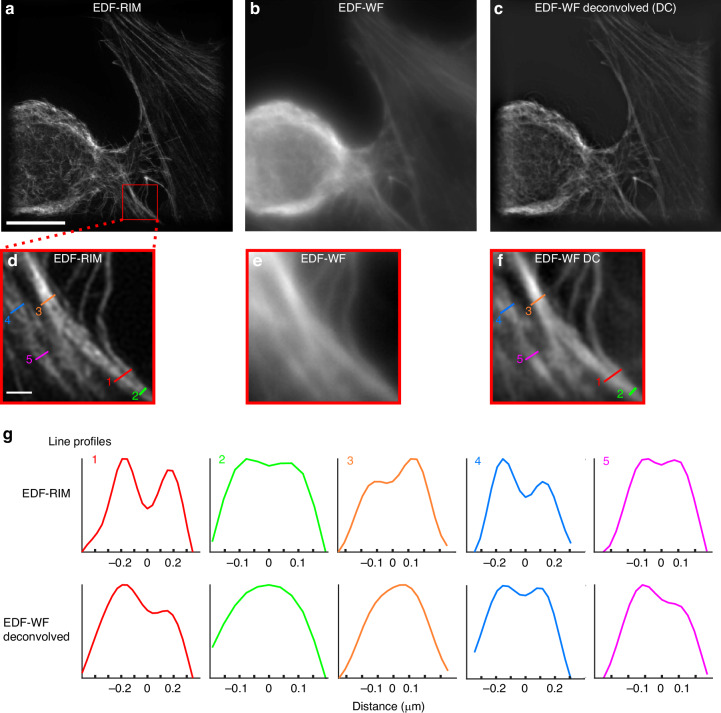
Fig. 2. Comparison of EDF-RIM with EDF-widefield.
a–f Phalloidin-alexa488 labeling of the actin cytoskeleton on a cultured cell imaged with EDF-RIM (a), EDF-WF (b) and EDF-WF deconvolved (c). The projected depth is 5.5 μm. Insets show a close-up view of the outlined region (d–f). Scale bar of the full image (a) and of the outlined region (d) are respectively 10 μm and 1 μm. g Line profiles along the segments shown in EDF-RIM (d) and deconvolved EDF-WF (f). EDF-RIM is able to distinguishes two filaments separated by 155 nm (lines 2,5) whereas deconvolved EDF-WF distinguishes at best two filaments separated by 269 nm (line 4)

