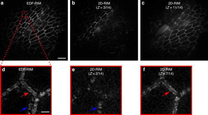
Fig. 3. Comparison of EDF-RIM with 2D-RIM.
a Image of desmosomes from the intestinal mouse epithelium with EDF-RIM, in which the entire 5.4 μm depth is acquired simultaneously. b, c Two slices (z = 3, and z = 11) of the same tissue imaged with 2D-RIM. The volume acquisition required 14 sequential z-slices. d Close-up view on the EDF-RIM image. e, f Close-up view on 2D-RIM at planes z = 2 and z = 7. Red and blue arrows point to structures in the EDF-RIM image that are only visible in one of the 2D-RIM images. Scale bar of the full image (a) and of the outlined region (d) are respectively 10 μm and 1 μm

