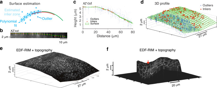
Fig. 4. Topographical estimation.
a–e Principle of topographical estimation illustrated on tight junctions from a chick neural tube (ZO-1 staining): one slice-by-slice stack is acquired for one speckled illumination. A consensual inlier/outlier classification of the bright points is then determined with RANSAC piece-wise fits (see “Methods”). Inliers are then interpolated. b Orthogonal view of the deconvolved slice by slice stack acquired for the surface estimation. c Orthogonal view of the surface, inliers and outliers for the chick neural tube data. d 3D view of surface, inliers and outliers. For sake of clarity, outliers (blue dots) are only shown on half of the surface. e The EDF-RIM image projected on the topography. f Projection of the EDF-RIM image of the desmosomes (Fig. 3a) on the estimated topography

