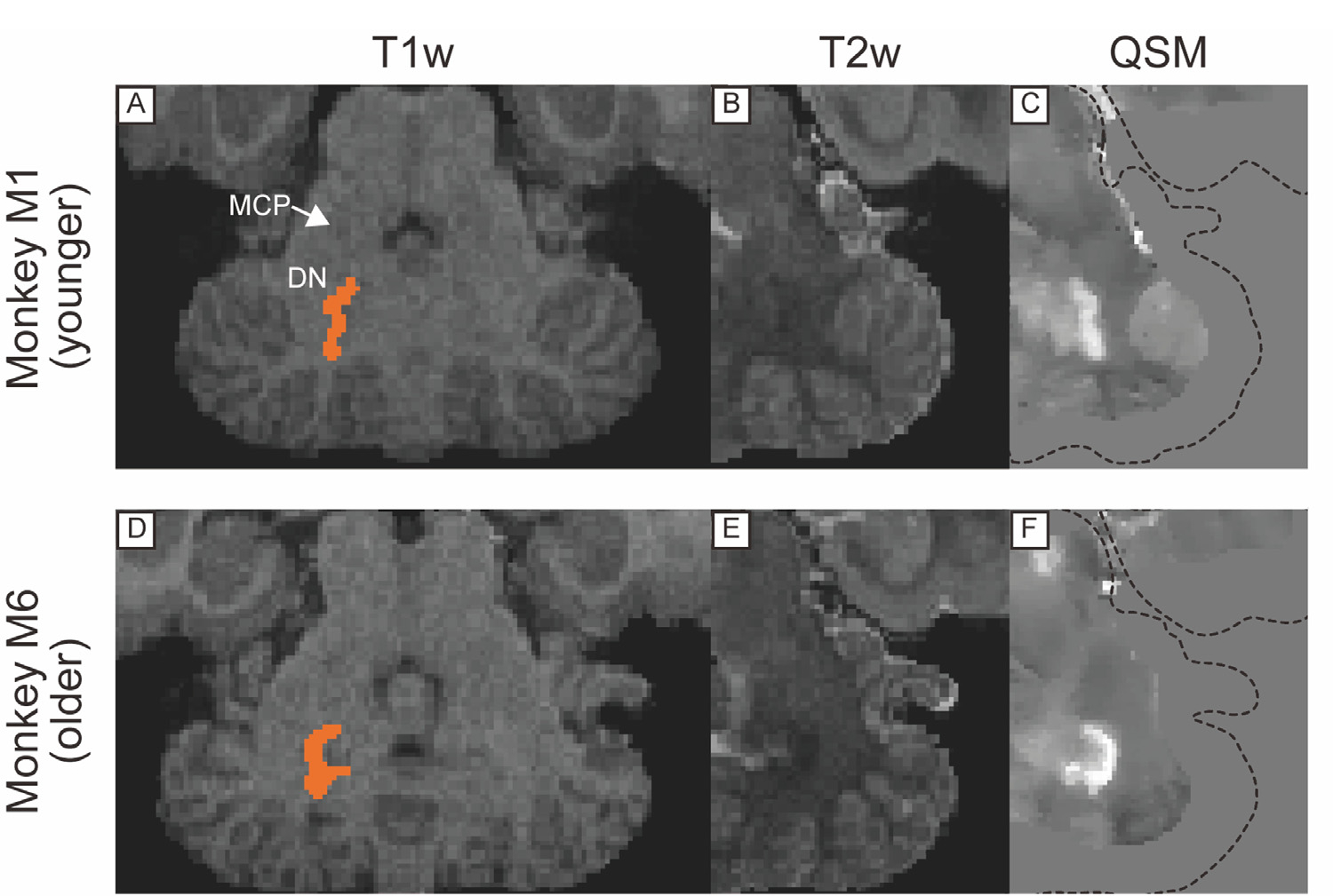
Fig. 5.

T1w, T2w, and QSM transverse images including dentate nucleus in cerebellum in a younger (A, B, C) and older monkeys (D, E, F). Dotted lines in (C) and (F) indicate the edge of the brain. Abbreviations: DN, dentate nucleus. MCP, middle cerebellar peduncle.
