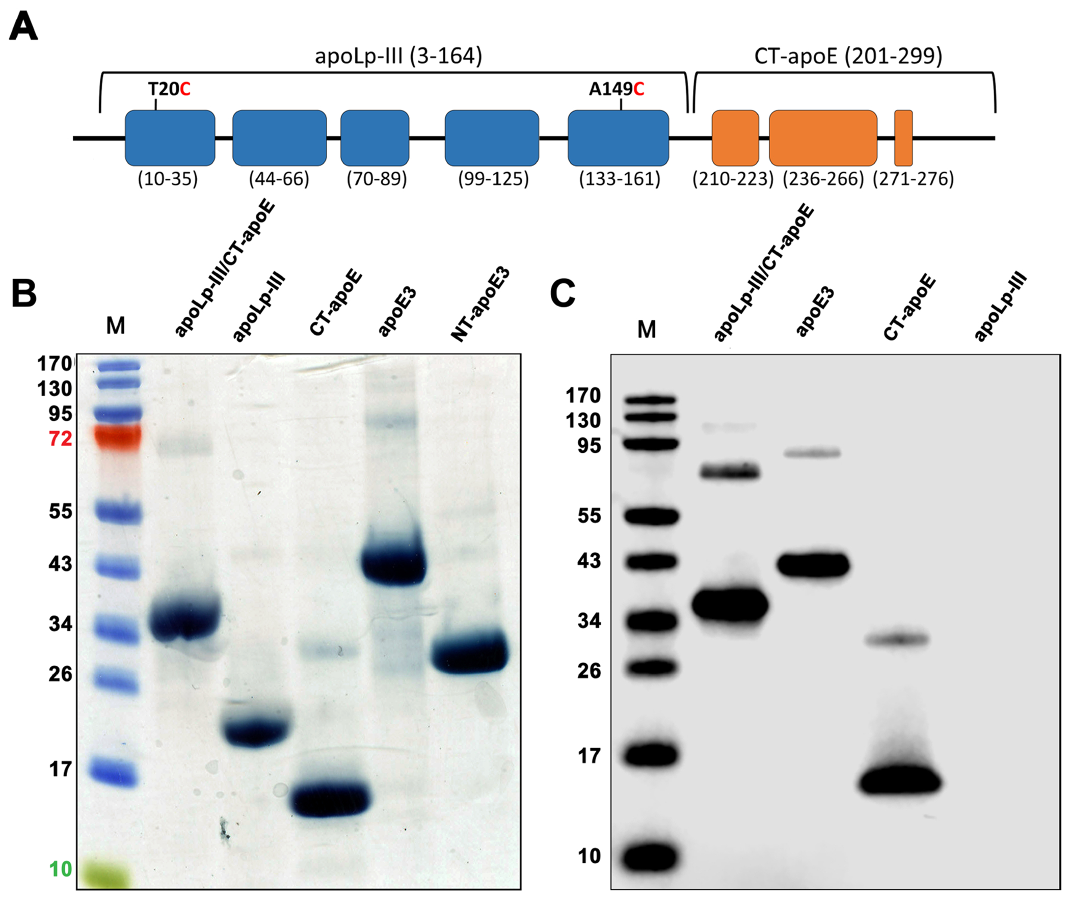
Figure 1.

ApoLp-III/CT-apoE chimera design and purification. Panel A: Schematic illustration of apoLp-III/CT-apoE chimera, with apoLp-III helices in blue and CT-apoE helices in orange. The chimera comprises residues 3-164 of L. migratoria apoLp-III and residues 201-299 of human apoE (helix boundaries indicated below each helix). For sake of convenience and clarity, residue numbering of the parent proteins was retained for the chimeras. Panel B: SDS-PAGE analysis of apoLp-III/CT-apoE chimera and relevant parent proteins under reducing conditions. Shown are the apoLp-III/CT-apoE chimera, apoLp-III, CT-apoE, apoE3, and NT-apoE3. The far-left lane (M) contains the molecular weight standards. Panel C: Western blot of purified recombinant proteins using a monoclonal antibody (3H1) specific for the CT domain of apoE3.
