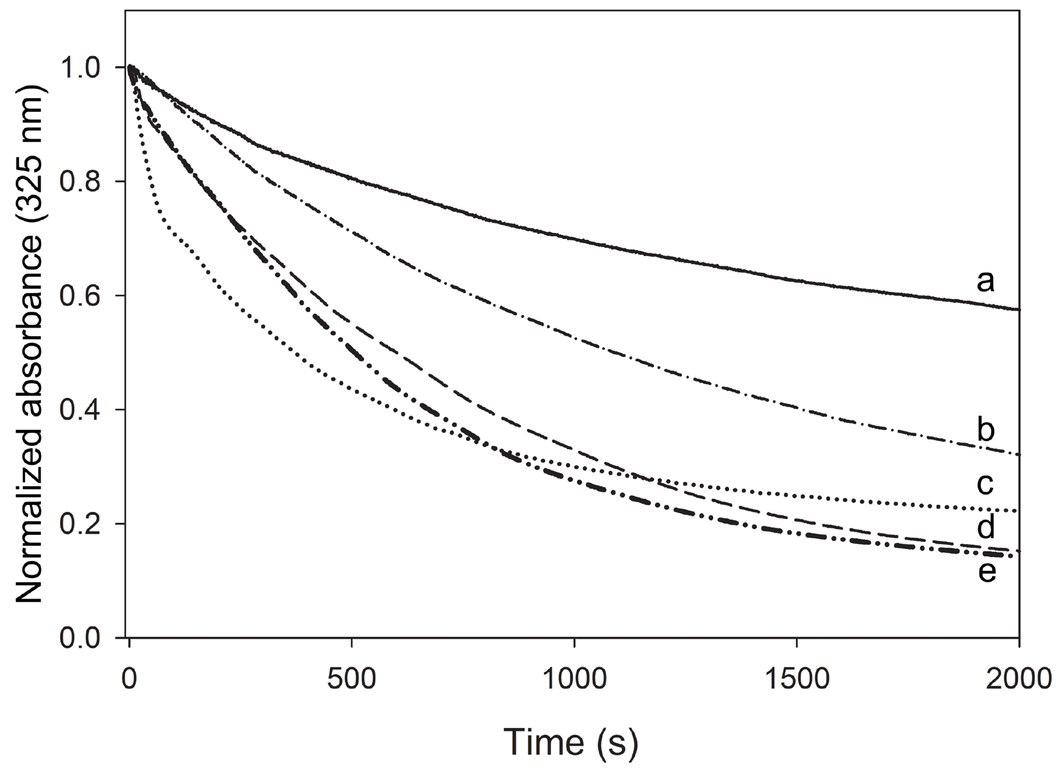
Figure 7.

Protein-induced solubilization of DMPC LUVs. Proteins and LUVs were mixed at a 1:1 weight ratio and incubated at 24.1 °C, during which LUVs were converted into small discoidal particles. Changes in LUV light scatter intensity were measured by following the absorbance at 325 nm. Shown are apoLp-III (a, solid line), NT-apoE3 (b, dash-dotted line), apoE3 (c, dotted line), apoLp-III/CT-apoE (d, dashed line), and CT-apoE (e, dash-double-dotted line). The samples did not contain DTT, therefore apoLp-III was mainly present in the oxidized state. When vesicles were monitored in the absence of protein, the absorbance did not change during the incubation time (not shown).
