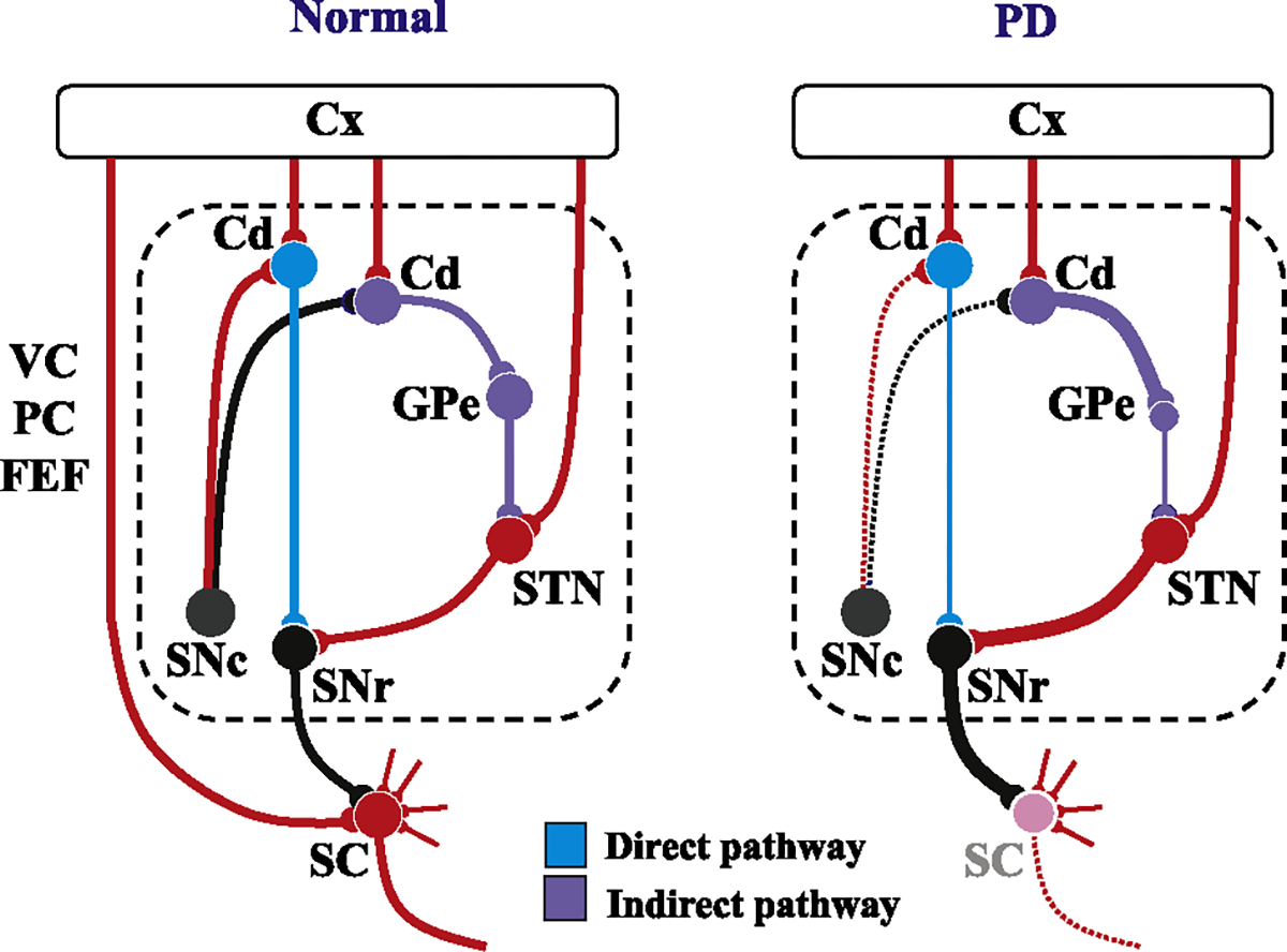
Fig. 2.

Schematic diagram showing the neural structures depicting the pathophysiology underlying saccade abnormalities in PD. Left figure: Schematic diagram depicting the neural structures involved in the control of saccades in a normal subject. Red lines indicate excitatory connections and black, blue, and purple lines indicate inhibitory connections. The dotted area designates the BG circuit. The blue curves denote the direct pathway and purple curves the indirect pathway (also see panel linked to the bottom of the figure). The pathways involved in the volitional control of saccades all converge onto the SC. Note that most of the inputs converging onto the SC are excitatory, while the only inhibitory input is from the basal ganglia (in the case of saccades, SNr). Some recent studies suggest the existence of direct projections from the prefrontal cortex to the brainstem saccade generator that play an important role in saccade inhibition, but these are not included in this figure. On the other hand, ascending inputs from the SNc to the caudate nucleus change the balance between the activity of the direct and indirect pathways of the BG circuit. Right figure: In PD, as the dopaminergic input from the SNc decreases, the balance between the activity of the direct and indirect pathways shifts in favor of the latter. Note that the inputs from the cortex is omitted in this figure. As a consequence, the SC becomes excessively suppressed compared to the SC in normal subjects (left figure), leading to the suppression of all kinds of saccades mediated by this structure. At the same time, the functioning of the direct pathway of the BG also decreases. These changes may explain why both VGS and MGS are affected in PD, and also why the degree of involvement is much stronger for the latter. Cd: caudate, Cx: the cerebral cortex, SC: superior colliculus, SNc: substantia nigra pars compacta, SNr: substantia nigra pars reticulata, GPe: external segment of the globus pallidus.
