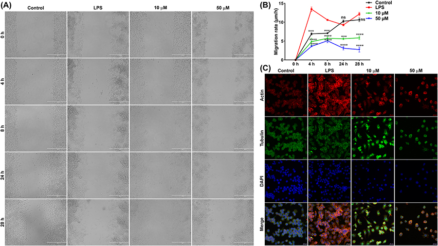
Fig. 2.
DJ-X-013 attenuated in vitro LPS-stimulated RAW 264.7 cell migration by regulating the expression of cytoskeletal proteins. (A) Representative phase contrast images show the closure of a scratch wound in a monolayer of LPS-stimulated RAW 264.7 macrophages in the presence or absence of DJ-X-013 over time, following incubation of 0, 4, 8, 24, and 28 h; scale bar 400 μm. (B) Plot showing that DJ-X-013 reduces cell migration rate. Statistical analysis was performed using one-way ANOVA followed by Dunnett’s post hoc test; n = 3. Data are presented as mean values ± SEM, ns p > 0.05; ***p < 0.001; ****p < 0.0001. (C) Representative immunofluorescence confocal microscopic images depict the alteration of actin and tubulin after treatment of LPS-stimulated RAW 264.7 macrophages with 10 or 50 μM DJ-X-013. Scale bar 20 μm; detected proteins (color): F-actin (red), tubulin (green), and DAPI for nucleus (blue). These results suggest that DJ-X-013 impedes the migration of macrophages and might modulate the polymerization or depolymerization of cytoskeletal proteins.

