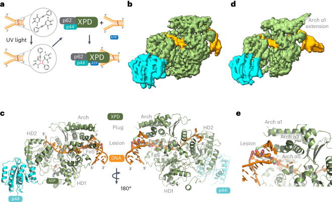
Fig. 1. Cryo-EM structure of the XPD complex in the presence of a Y-forked DNA substrate containing an engineered cross-link.
a, Schematic description of sample preparation prior to vitrification. b, Cryo-EM map of the class 1 XPD–p44–DNA complex. XPD is colored in green, p44 is colored in cyan and DNA is colored in orange. c, Left, structural model of the XPD–p44–DNA complex in cartoon representation, color-coded as in b. Right, Rotation (180°) of the model around its y axis. d, Cryo-EM map of the class 2 XPD–p44–DNA complex, color-coded as in b. e, Close-up of the dsDNA–ssDNA junction and the cross-link at the Arch domain of XPD, color-coded as in b. The cross-link is shown as spheres.

