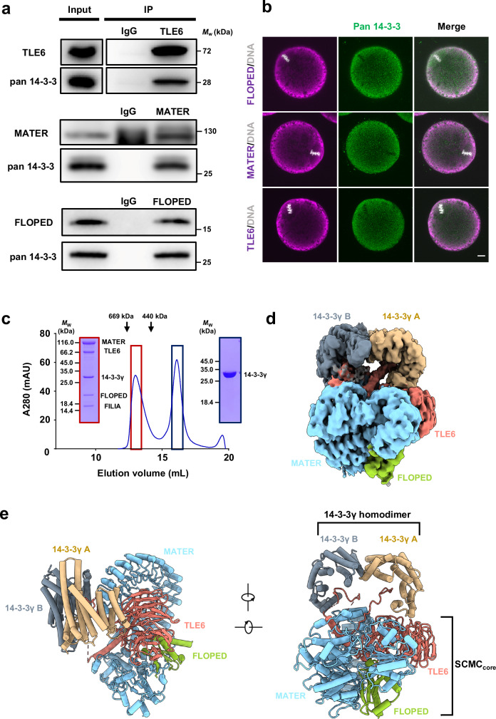
Fig. 1. 14-3-3 exhibits as an SCMC component.
a Normal oocyte lysates, before (Input) or after immunoprecipitation with antibody to TLE6 (top), MATER (middle) or FLOPED (bottom) were immunoblotted and probed with antibodies to pan 14-3-3 and TLE6, MATER, or FLOPED, respectively. IgG, normal immunoglobulin (negative control). b Normal oocytes isolated from normal females were fixed, permeabilized, and incubated with antibodies to pan 14-3-3 and FLOPED (top), MATER (middle), or TLE6 (bottom), and with DAPI to visualize DNA. The oocytes were imaged by confocal microscopy. Scale bar, 10 μm. Colocalization of pan 14-3-3 and FLOPED, MATER, or TLE6 was presented in the merge. c In vitro reconstitution of mouse SCMC-14-3-3γ. The mouse SCMC quaternary complex and 14-3-3γ protein were incubated in lysis buffer. Size-exclusion chromatography (Superose™ 6 Increase 10/300 GL) was performed to separate the SCMC-14-3-3γ (marked in dark red box) and excess 14-3-3γ (marked in dark blue box). The SCMC-14-3-3γ is composed of His-tagged MATER (1–1059 aa), Strep-tagged TLE6 (48–581 aa), Strep-tagged FLOPED (1–164 aa), Strep-tagged FILIA (1–124 aa), and 14-3-3γ (1–247 aa). The column was calibrated with thyroglobulin (669 kDa) and ferritin (440 kDa). d Cryo-EM map of the SCMC-14-3-3γ. 14-3-3γ A and 14-3-3γ B refer to two protomers in 14-3-3γ homodimer. MATER, TLE6, and FLOPED are colored in light blue, salmon, and yellow green, respectively. The homodimer of 14-3-3γ is distinguished by 14-3-3γ A (burlywood), which is proximal to the WD40 repeat domain of TLE6, and 14-3-3γ B (gray), which is close to the leucine-rich repeat domain of MATER. The map was shown at level 0.00679. e Two views of the cartoon presentation of the SCMC-14-3-3γ structure. The color scheme is consistent with panel d. The SCMCcore indicates the core complex consisting of MATER, TLE6, and FLOPED.

