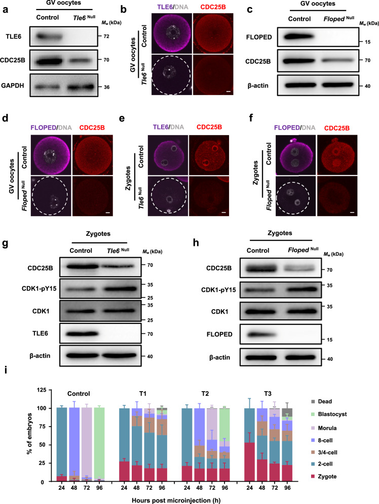
Fig. 5. The SCMC-14-3-3 promotes the stability of CDC25B for mitotic entry during the maternal-to-embryo transition.
a Immunoblotting of CDC25B in normal control and Tle6Null germinal vesicle (GV) oocytes. b Immunofluorescence (IF) staining of CDC25B in GV oocytes isolated from control or Tle6Null female mice. Scale bar, 10 μm. c Immunoblotting of CDC25B in control and FlopedNull GV oocytes. d IF staining of CDC25B in GV oocytes isolated from control or FlopedNull female mice. Scale bar, 10 μm. e, f IF staining of CDC25B in zygotes from control and Tle6Null (e) or FlopedNull (f) female mice. Zygotes were flushed from the oviduct of female mice that underwent superovulation with gonadotrophins and were mated with normal males. DNA was visualized with DAPI. Scale bar, 10 μm. g, h Immunoblotting of CDC25B, CDK1, and CDK1-pY15 in zygotes from control and Tle6Null (g) or FlopedNull (h) female mice. Zygotes were flushed from the oviduct of female mice that underwent superovulation with gonadotrophins and were mated with normal males. pY, phosphorylated tyrosine. i, Mouse zygotes were cultured for 24 h, 48 h, 72 h, and 96 h after co-microinjection with mCherry-Trim21 mRNA + isotype IgG + mRNA solvent (control), mCherry-Trim21 mRNA + anti-TLE6 antibody + mRNA solvent (T1), mCherry-Trim21 mRNA + anti-TLE6 antibody + Cdc25b mRNA (T2), or mCherry-Trim21 mRNA + anti-TLE6 antibody + Cdc25bC483A/E484A mRNA (T3). The proportions of embryos on different stages were calculated. N = 3 independent experiments. The data are presented as mean ± S.D. (standard deviation).

