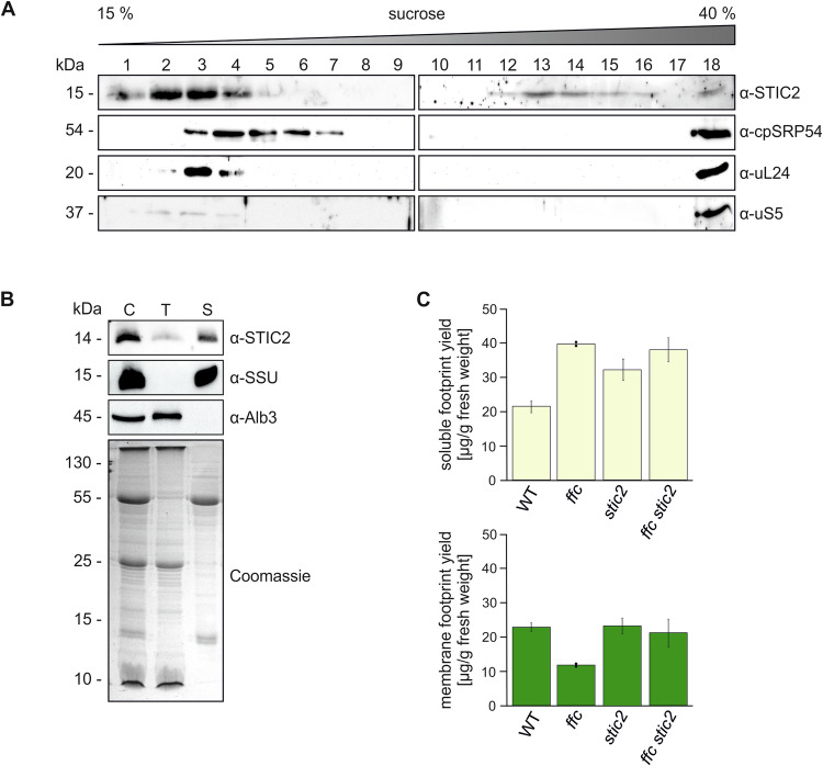
Figure 4. Analysis of cofractionation of STIC2 with stromal ribosomes and thylakoid membranes and the effect of the stic2 mutation on ribosomal footprints.
(A) The interaction of endogenous STIC2 and ribosomes was tested by sucrose density gradient centrifugation of leaf extracts of A. thaliana WT plants. The gradient fractions were separated by SDS-PAGE and applied to immunoblotting using the indicated antibodies. A representative experiment from two independent biological and two technical replicates is shown. (B) Isolated chloroplasts from A. thaliana WT were lysed and separated into stromal and thylakoid fractions by centrifugation. Samples equivalent to 5 µg chlorophyll of chloroplast extract (C), washed thylakoids (T), and stroma (S) were separated on SDS-PAGE and blotted for immunodetection using antisera raised against STIC2, Alb3 and the small subunit of Rubisco (SSU). The Alb3 insertase and SSU were used as controls for successful fractionation. A Coomassie blue stained SDS gel served as loading control. A representative experiment from two independent biological replicates is shown. (C) Ribosome footprint yield was determined for soluble (yellow) and membrane (green) fractions of WT, stic2-3, ffc1-2, and ffc1-2 stic2-3 and normalized to the amount of fresh weight used as starting material. Means and SDs are derived from two (ffc1-2) or three (WT, stic2-3, ffc1-2 stic2-3) independent biological replicates. Source data are available online for this figure.

