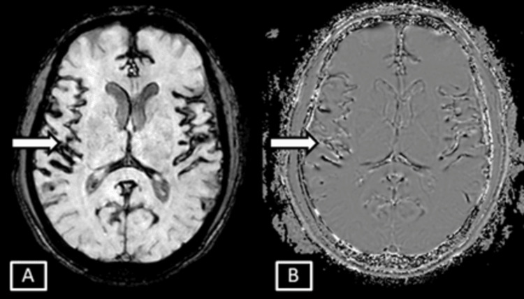
Figure 6. Case 2 MRI brain axial SWI magnitude (A) and phase (B) sequences.
Images show symmetrical SWI hypointensites seen involving the sulcal spaces along the cortical surface of bilateral cerebral hemispheres and along the falx represented by the white arrows.
SWI: Susceptibility Weighted Imaging

