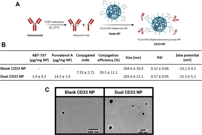
Figure 5.
Preparation and characterization of CD33 NPs. (A) Schematic overview of the antibody–nanoparticle conjugation process, using maleimide–thiol chemistry. (B) Table summarizing conjugated NPs characteristics in terms of drug loading, amount of antibody conjugated obtained via Micro BCA, DLS-measured hydrodynamic diameter (nm), and polydispersity index (PdI) values, and PALS-measured zeta potential values. Data are presented as mean ± SD, from measurements performed in triplicate and averaged from at least n = 3. (C) TEM images of CD33 NPs, representative of two independent experiments.

