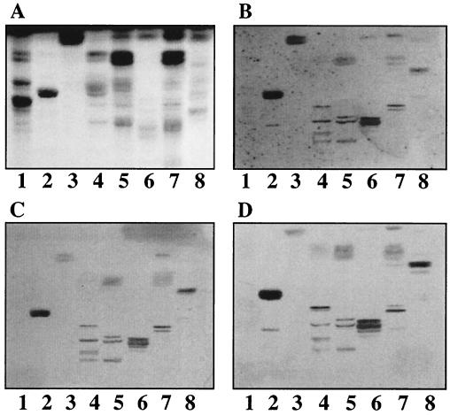
FIG. 2.
Binding of 125I-labeled SA11 and NCDV rotaviruses to mixtures of nonacid glycosphingolipids on TLC plates. Glycosphingolipids were separated on TLC plates using chloroform-methanol-water (60:35:8 by volume). One chromatogram (A) was stained with anisaldehyde. Other chromatograms were incubated with the following purified radiolabeled viral particles: TLP from SA11 (B), DLP from SA11 (C), and DLP from NCDV (D). Each lane contained 40 μg of glycosphingolipid mixture. Lanes 1, nonacid glycosphingolipids of human erythrocytes, blood group AB; lanes 2, nonacid glycosphingolipids of guinea pig erythrocytes; lanes 3, nonacid glycosphingolipids of bovine intestine; lanes 4, nonacid glycosphingolipids of bovine erythrocytes; lanes 5, partially purified nonacid glycosphingolipids of bovine erythrocytes; lanes 6, nonacid glycosphingolipids of mouse small intestine; lanes 7, nonacid glycosphingolipids of human large intestinal adenocarcinoma; lanes 8, nonacid glycosphingolipids of guinea pig small intestine.

