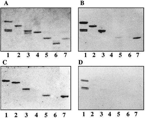
FIG. 5.
Binding of 125I-labeled SA11 and NCDV rotaviruses to pure gangliosides on TLC plates. Glycosphingolipids were separated on TLC plates using chloroform–methanol–0.25% KCl in water (50:40:10 by volume). One chromatogram (A) was stained with anisaldehyde. Other chromatograms were incubated with the following purified radiolabeled viral particles: TLP from SA11 (B), TLP from NCDV (C), and DLP from SA11 (D). Each lane contained 2 μg of pure glycosphingolipids. Lanes 1, GA2 and/or GA1; lanes 2, NeuGc-GM3; lanes 3, NeuGc-GM2+NeuGc-GM1 [GalNAcβ4(NeuGcα3)Galβ4Glcβ1Cer plus Galβ3GalNAcβ4(NeuGcα3)Galβ4Glcβ1Cer]; lanes 4, NeuAc-GM1; lanes 5, NeuAc-GD1a; lanes 6, NeuAc-GD1b; lanes 7, NeuGc-GD1a.

